Cervical spine special tests
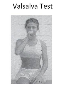
Valsalva test
- Procedure: The patient is seated with the thumb in the mouth and attempts to push the thumb out by blowing out hard.
- Assessment: The pushing increases the intraspinal pressure, revealing the presence of space-occupying masses such as extruded intervertebral disks, tumors, narrowing due to osteophytes, and soft tissue swelling. This leads to radicular symptoms entirely confined to the respective dermatome or dermatomes.
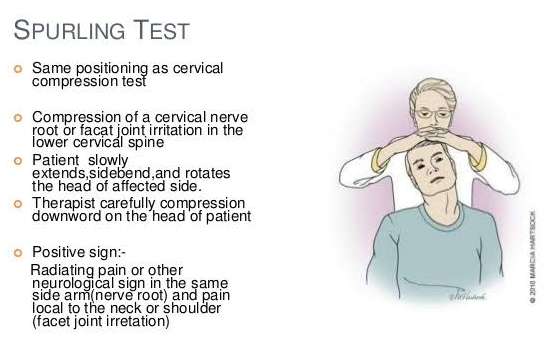
Spurling test ( Foraminal compression test )
- Assesses facet joint pain and nerve root irritation.
- Procedure: The patient is seated with the head rotated and tilted to one side. The examiner stands behind the patient with one hand placed on the patient’s head. With the other hand, the examiner lightly taps (compresses) the hand resting on the patient’s head. If the patient tolerates this initial step of the test, it is then repeated with the cervical spine extended as well.
- Assessment:This test provides clinical evidence of both a facet syn- drome and nerve root compression. Where facet joint irritation or nerve root compression is present, the examination will intensify the pain. Simultaneous extension of the cervical spine narrows the intervertebral foramina by 20–30%. Existing radicular pain will be increased by this movement.
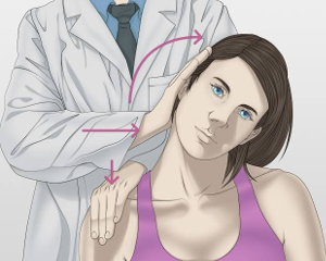
Shoulder depression test
- Assess the nerve root compression
- Procedure: Side Flex patient’s head on unaffected side then apply a downward pressure on the opposite shoulder (affected side).
- Positive sign- increase in pain.
- This Test provides evidence for nerve root compression.
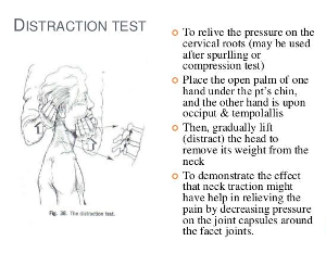
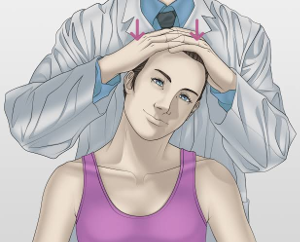
Distraction test
- Differentiates between radicular pain in the back of the neck, shoulder, and arm and ligamentous or muscular pain in these regions.
- Procedure: The patient is seated. The examiner grasps the patient's head about the jaw and the back of the head and applies superior axial traction.
- Assessment: Distraction of the cervical spine reduces the load on the intervertebral disks and exiting nerve roots within the affected levels or segments while producing a gliding motion in the facet joints. Reduction of radicular symptoms, even in passive rotation, when the cervical spine is distracted is a sign of discogenic nerve root irritation. Increased pain during distraction and rotation suggests a functional impairment in the cervical spine due to muscular or ligamentous pathology.
Percussion Test
- Procedure: With the patient’s cervical spine slightly flexed, the exam- iner taps the spinous processes of all the exposed vertebrae.
- Assessment: Localized nonradicular pain is a sign of a fracture or of muscular or ligamentous functional impairment. Radicular symptoms indicate intervertebral disk pathology with nerve root irritation.
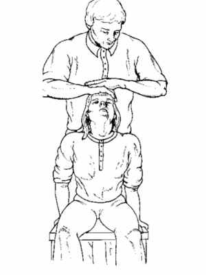
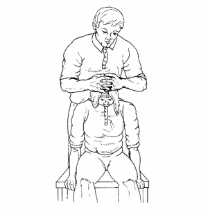
Jackson Compression Test
- Procedure: The patient is seated. The examiner stands behind the patient with his or her hand on the top of the patient’s head and passively tilts the head to either side. In maximum lateral bending, the examiner presses down on the head to exert axial pressure on the spine.
- Assessment: The axial loading results in increased compression of the intervertebral disks, exiting nerve roots, and facet joints. Pressure on the intervertebral foramina acts on the facet joints to elicit distal pain that does not exactly follow identifiable segmental dermatomes. Presence of nerve root irritation will cause radicular pain symptoms.
Flexion Compression Test
- Procedure: The patient is seated. The examiner stands behind the patient and passively moves the cervical spine into flexion (tilts the patient’s head forward). Then axial compression is applied to the top of the head.
- Assessment: This is a good test of the integrity of the intervertebral disk. In the presence of a posterolateral disk extrusion, this maneuver will press the extruded portion of the disk in a posterior direction, resulting in increasing compression of the nerve root. An increase in radicular symptoms can therefore indicate the presence of a postero- lateral disk extrusion.
Extension compression test
- Procedure: The patient is seated and the examiner stands behind the patient. The cervical spine is extended 30°. The examiner then applies axial compression to the top of the head.
- Assessment: This test assesses the integrity of the intervertebral disk. Where a posterolateral extrusion with an intact annulus fibrosus is present, shifting the pressure on the disks anteriorly will reduce symp- toms. Increased pain without radicular symptoms usually indicates an irritation in the facet joints as a result of decreased mobility due to degenerative changes.